

2026-11-06
Can Veneers Change Your Patient's Bite? Understanding Anterior Guidance
Written by Dr. Agatha Bis
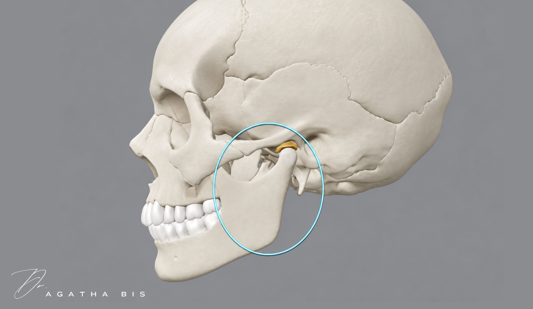
A patient wants longer anterior teeth. You plan the new incisal edges, improve the proportions, and create a beautiful smile.
But have you considered where those new incisal edges will stop the mandible?
This is a question I believe deserves more attention in cosmetic dentistry because anterior restorations influence more than appearance.
The Teeth Determine Where the Mandible Closes
The teeth provide the contact stops that guide the mandible into maximum intercuspation. Changing those stops can alter the pathway of closure and the position of the condyles within the joints.
When we lengthen anterior teeth or increase the thickness of their lingual contours, we may change how the lower incisors contact the upper teeth.
If the new restorations occupy space that the mandible requires during closure or function, the patient may experience restrictive contacts or a change in mandibular movement.
That does not mean every anterior restoration causes TMJ problems. It means we need to understand the patient's existing functional relationships before changing them.
Why Anterior Guidance Matters
Anterior guidance influences mandibular movements and the way teeth separate during excursions.
Williamson and Lundquist demonstrated that immediate posterior disclusion through anterior guidance reduced elevator muscle activity during excursive movements [1].
This relationship matters because a restoration that changes guidance also changes how forces are distributed during movement.
Consider a patient with significant anterior wear. Restoring the teeth to their original length may seem straightforward, but dentoalveolar compensation may have maintained vertical dimension as the teeth shortened.
The restorative space that once existed may no longer be available.
If we rebuild the anatomy without assessing the closing path and functional envelope, we may introduce a restriction that was not present before treatment.
What About Condylar Position?
In my clinical framework, I examine how changes in anterior stops may influence mandibular positioning and loading within the TMJ.
Imaging studies have reported associations between posterior condylar position and disc displacement [2,3]. However, condylar position alone is not diagnostic, and the relationship between occlusion and TMD is complex.
This is why I evaluate the individual patient rather than making treatment decisions from an occlusal feature or radiographic finding in isolation.
I want to know whether the closing position is reproducible, whether the patient has joint or muscle symptoms, and whether the proposed restorations will maintain comfortable function.
Test the Design Before Making It Permanent
Digital smile design allows us to visualize esthetics, but it cannot fully test function.
When meaningful changes in incisal position or guidance are planned, provisional restorations allow us to assess overbite, overjet, speech, mandibular movement, muscle comfort, and closure before definitive ceramic placement.
We should not expect the patient to adapt to a final restoration when we had the opportunity to evaluate the proposed design first.
I explore this relationship between anterior restorations, condylar position, and functional stability in my article, Cosmetic Dentistry on an Unstable Foundation.
Learn to Connect Occlusion with Treatment Planning
In Hands-On Level 1, we establish the diagnostic foundation through comprehensive examination.
In Hands-On Level 2: Occlusion and Treatment Planning, we focus on understanding functional relationships, force distribution, and how those findings guide restorative decisions.
Explore Hands-On Level 1 | Explore Hands-On Level 2
References
Williamson EH, Lundquist DO. Anterior guidance: its effect on electromyographic activity of the temporal and masseter muscles. J Prosthet Dent. 1983;49(6):816–823.
Gateno J, et al. A comparative assessment of mandibular condylar position in patients with anterior disc displacement. J Oral Maxillofac Surg. 2004;62(1):39–43.
Bonilla-Aragon H, et al. Condyle position as a predictor of temporomandibular joint internal derangement. J Prosthet Dent. 1999;82(2):205–208.
Manfredini D, et al. Temporomandibular disorders and dental occlusion. J Oral Rehabil. 2017;44(11):908–923.